Customer Case Study
VU University Medical Center (VUmc) & Academic Medical Center (AMC), Amsterdam
Connected images
The ‘new normal’
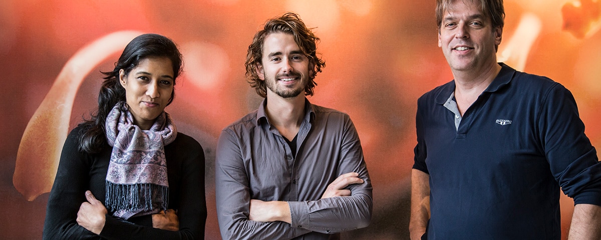
Varsha Birdja
Application specialist
Application specialist
Project leader Image Management
A centralized, truly enterprise-wide image management strategy on the firm foundation of Enterprise Imaging
A shared image management strategy
Two academic hospitals in Amsterdam – VUmc and AMC – are collaborating with the intention to create an alliance that will result in combined facilities totaling 1,700 beds and 13,000 staff (FTE).
A common strategy for image management, implemented in collaboration with AGFA HealthCare and based on the Enterprise Imaging platform, is a key factor of this merger.
“Imagine hundreds of clinicians spending half an hour, every day, to look for their images, transport them and have them available in their viewer. Today, those clinicians immediately have access to images going back even 10 or 15 years, with our Enterprise Imaging solution. Think how much time and effort that saves!
And already, it has become the ‘new normal’ for them.”
“This remarkable situation is the result of an extensive project to implement Enterprise Imaging in parallel with an electronic medical record (EMR) at the hospitals.”
VUMC and AMC’s image management vision is founded on 4 pillars
Streamlined and standardized workflows for image capture and acquisition
Centralized image archiving and back-up of all images
A single image viewer for all types of images, integrated in the EMR
Centralized exchange of image data
An end-of-life archive and multiple pacs
VUmc and AMC were facing a number of image management challenges. In 1996, VUmc had built its own image archive, which was now coming to its end of life. At the same time, both hospitals had a number of different departmental PACS and caregivers were not able to retrieve images from other departments. Finally, as Ernest describes, “more and more departments in hospitals are generating images.”
To address these issues, he explains, VUmc and AMC decided to increase centralization. “We envisaged a centralized enterprise imaging platform. But we didn’t want to replace all the specialty IT systems in each department; instead we wanted to connect them as much as possible to the centralized platform. That way, users could keep their front-end applications, but the data would be available to the entire organization.”
Critically, the platform also had to integrate with the EMR chosen by both VUmc and AMC. “We were planning to introduce an enterprise-wide EMR as well, to provide one patient record with all available data. We wanted images – regardless of their source – to fundamentally be part of this.”
Together, the hospitals developed the detailed European tender for an enterprise imaging solution, which took more than a year. “We included questions on the system’s functionality and the license cost, of course, but also about support from the vendor, the ICT infrastructure needed, the composition of our own team that would be needed for the project, etc. And we visited other hospitals as well as the vendors. In the end, AGFA HealthCare’s offer had the best results!”
“The basic idea is to work more efficiently, to deliver reliable care and to decrease errors. Having all the images available on one system for multidisciplinary meetings and boards offers a great advantage in attaining good patient outcomes, too. And finally, with centrally available images, we reduce duplicated exams.”
A diverse team: technical knowledge and communication skills
With the Enterprise Imaging platform selected, the implementation project could begin. VUmc and AMC began with designing the hardware infrastructure and preparing the data migration.
While AMC opted to start off with the EMR and the imaging archive, VUmc chose to roll out Enterprise Imaging and the EMR simultaneously, half a year after AMC went live with the EMR. “This approach required a lot of parallel work between the Image Management team and the EMR team,” explains Ernest.
“We set up a functional management group for image management, with people who are both technically skilled and who can easily communicate with users in the hospital. Our group is part of the EVA service center (the joint service center for both hospitals), which is dealing with the functional management of the entire medical record, including images.”
Mapping modalities, defining workflows
With images coming from so many domains of the patient’s care, there is a great diversity of formats that must be managed, stored and accessible in the Enterprise Imaging platform.
“When we are preparing to connect a department, we talk with the people to inventory the kinds of exams they perform, how the department works and what their needs are. This can be very precise, for example the order in which they want to see the images from a specific study. We check whether the exams are being requested and who is requesting them, and how the modality stores the images.
“Departments work in different ways and use different modalities: some support DICOM and DICOM worklists, others only support jpg, or can only print,”
Agfa HealthCare’s Solution
The Enterprise Imaging solution, being rolled out enterprise-wide at VUmc, includes the following components:
- The Enterprise Imaging VNA (Vendor Neutral Archive) consolidates all imaging data from multiple systems, departments, facilities and vendors, into a central clinical data foundation.
- Enterprise Imaging Exchange allows fast, secure, reliable transfer of any and all patient studies, with no CDs or DVDs.
- The web-based XERO Universal Viewer provides secure access to DICOM and non-DICOM imaging data from different departments and multiple sources, in one view, to anyone inside and outside of the hospital who needs it.
This workflow-centric platform can be adopted step-by-step, at the hospital’s own speed.
Then we decide how to archive the modality’s images in DICOM, we create procedure codes and we give our colleagues in the department demos and training.” “It isn’t always easy to find the right contact persons for each department,” adds Varsha Birdja, application specialist, Image Management. “There can be a different person for each modality.”
The group defined eight generic workflows; each device is mapped to one of these. “When we go into a new department, this helps us determine how to proceed and what to ask. And when we have implemented a specific workflow in one department, we can then reuse it in another,” Varsha continues. The workflow for uploading CDs is also a key attention point for the hospitals. “Our hospitals receive about 60,000 CDs with images each year (whether sent from referring hospitals or brought by patients), which is about 15% of our total volume of images,” comments Varsha.
“So, this is a very important workflow!” “And some hospitals supply images via third-party digital systems; those have to connect to our system, too,” adds Toon. This workflow has already been implemented in VUmc, and will soon be implemented in AMC.
Finally, there is a special workflow for ‘confidential’ images. “These include images such as those from pediatric psychology: images of children that are thought to have been abused or other very sensitive situations,” highlights Ernest. “The images are kept in the archive, but are only accessible by specific users.
And they are not available through the EMR, but directly via the XERO Viewer.”
Migrating 20 years of images
Already, more than 200 devices have been connected, and the number rises each week, says Ernest. The previous DICOM archive, with 20 years of images, has also been migrated into the Enterprise Imaging platform: “about 4.5 million studies, representing almost half a billion images mainly from radiology, but also from other departments, such as obstetrics/ gynecology.”
One challenge that arose when migrating the exams was ensuring that all studies have the same names. Ernest notes that for some studies, up to 50 different names had been used over the past decades. “We mapped all those different names to one generic study name and procedure code, that is also used in the EMR: instead of thousands of codes, we now have less than 1000 EMR codes. So, all relevant prior studies can be easily retrieved.”
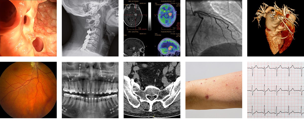
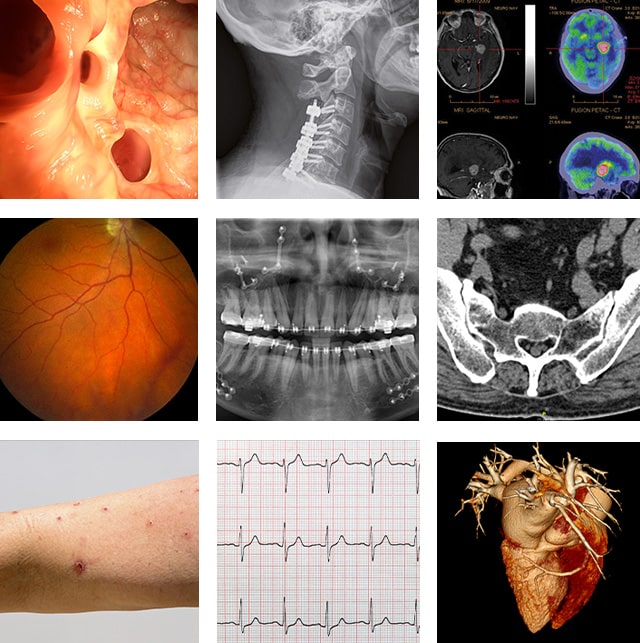
Supported images include:
Anesthesiology, Cardiac Surgery, Dermatology, Gynecology, Gastroenterology, Intensive Care, Pediatric surgery, Nephrology, Neurology, ENT, Pulmonology, Oncology, Orthopedics, Plastic surgery, Radiotherapy, Urology, Blood vessal lab, Obstetrics, …
Efficiency and compromise: a delicate balance
The team started by connecting the ENT (ear, nose and throat), ophthalmology and gastroenterology units, and the vascular lab. Varsha explains, “These departments all had paper-based workflows, which we wanted to eliminate, so they had the highest priority.
Next we connected the radiology images to the VNA, obstetrics and gynecology, cardiology, pulmonary diseases, emergency care, intensive care and pediatrics.” While the new workflows require users to adapt some of their work habits, the team also strives to find ways to compromise.
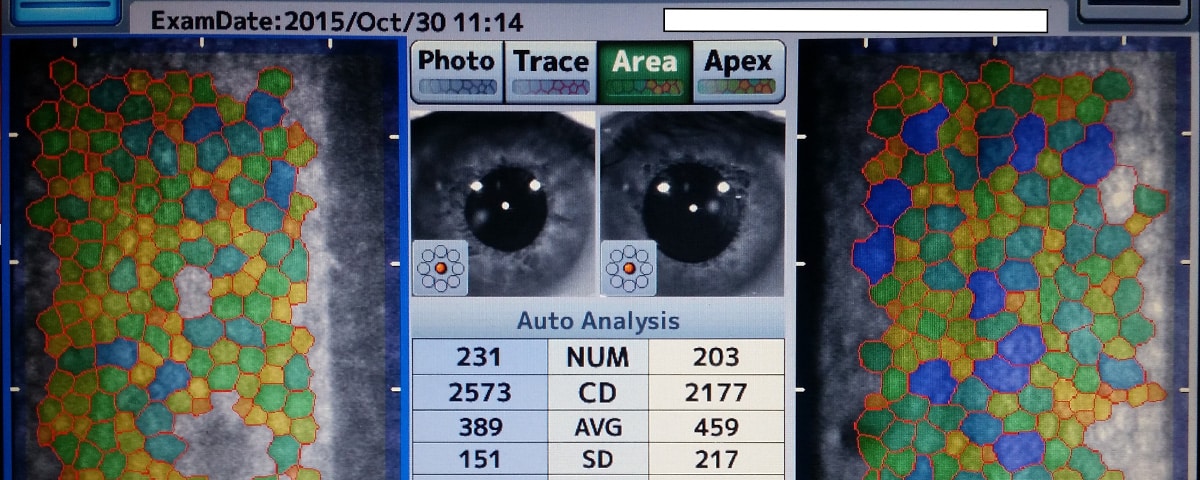
Toon comments on the issue of exam ordering, for example: “Ophthalmology is now working with requests (or orders), which it didn’t do in the past; the orders are displayed in the modality worklist.
But for gynecology, we know that 95% of all pregnant women visiting the department get an ultrasound. So, based on the type of visit scheduled, we automatically create an order in the background for an ultrasound. If it isn’t performed, it is removed from the worklist at the end of the day, and after five or six days, the order disappears from the system.”
Evolving modalities and devices
The number of modalities to connect continues to grow, though, and the teams have defined procedures for adding them.
“OCT (optical coherence technology), originally used in ophthalmology, is now being used in cardiology as well, so we will connect two OCTs in cardiology. And when we acquire new devices, a crossdepartmental process in the two hospitals makes sure the purchased modalities can be easily connected to the Enterprise Imaging solution.”
AGFA HealthCare’s support has been very important to the success of the project, the colleagues agree: “When we started designing the workflows, we usually had one or two AGFA HealthCare people in our hospital for functional support.
They helped us with migrating the old image archive, which was extremely complex and extensive, and with integrating the Enterprise Imaging platform with the EMR, as well as delivering training to the key users.
We continue to receive on-site support from AGFA HealthCare about one day every two weeks, to help solve current issues and configure new things,” explains Ernest.
Delivering reliable care and reducing errors
There are now around 5,000 users of the system at VUmc, and AMC is getting ready to roll-out the Enterprise Imaging solution as well. “In general, most people find it easy to use, although it was easier for certain users or modalities to make the switch than for others,” comments Varsha.
Toon agrees: “Overall, everybody is happy that the data is digitized and centrally available through the EMR.
The basic idea is to work more efficiently, to deliver reliable care and to decrease errors. Having all the images available on one system for multidisciplinary meetings and boards offers a great advantage in attaining good patient outcomes, too. And finally, with centrally available images, we reduce duplicated exams.
We have also sped up certain exams, for example in ophthalmology a specific modality transferred images of each eye individually, which took quite a while. We adapted the system to transfer the images of the two eyes simultaneously, saving time.”
He concludes, “What I like most about my job now is that I know we are working on something that is very important to the hospital, and ultimately, the patients.”
Use cases
Intubation videos enhance patient safety
Intubating patients under narcosis can be very difficult, and placing a camera on top of the intubation tube can be helpful.
Various departments create these intubation videos.
The systems will soon be connected to the Enterprise Imaging platform enabling the videos to be stored, so that the physician can review them when a patient returns for surgery.
Collaboration reduces duplicate exams
Stroke patients in Amsterdam are cared for at AMC. When a CT shows that a patient has had a stroke, he is transferred to AMC; timely delivery of the CT results at the receiving hospital means the care team does not have to duplicate exams.
The Enterprise Imaging platform supports this type of collaboration between hospitals, including via the CD workflow.
Enterprise Imaging also supports collaboration between colleagues in different departments. To give one example, pediatric cardiologists can now easily view ultrasounds made in the gynecology / obstetrics department.
Reducing duplicate exams through collaboration saves costs, saves time and enhances the patient’s experience.
Sharing ECG workflows across departments
Before the introduction of Enterprise Imaging, ECGs from the various departments were paperbased (except for cardiology, which stored them on an old IT system).
This prevented them from being shared between colleagues in different specialties.
Now, all ECG systems from the entire hospital are in the process of being connected to the centralized Enterprise Imaging platform and the ECGs are in digital format, making them easy to share. Cardiologists can view ECGs from clinical departments, and vice-versa.
